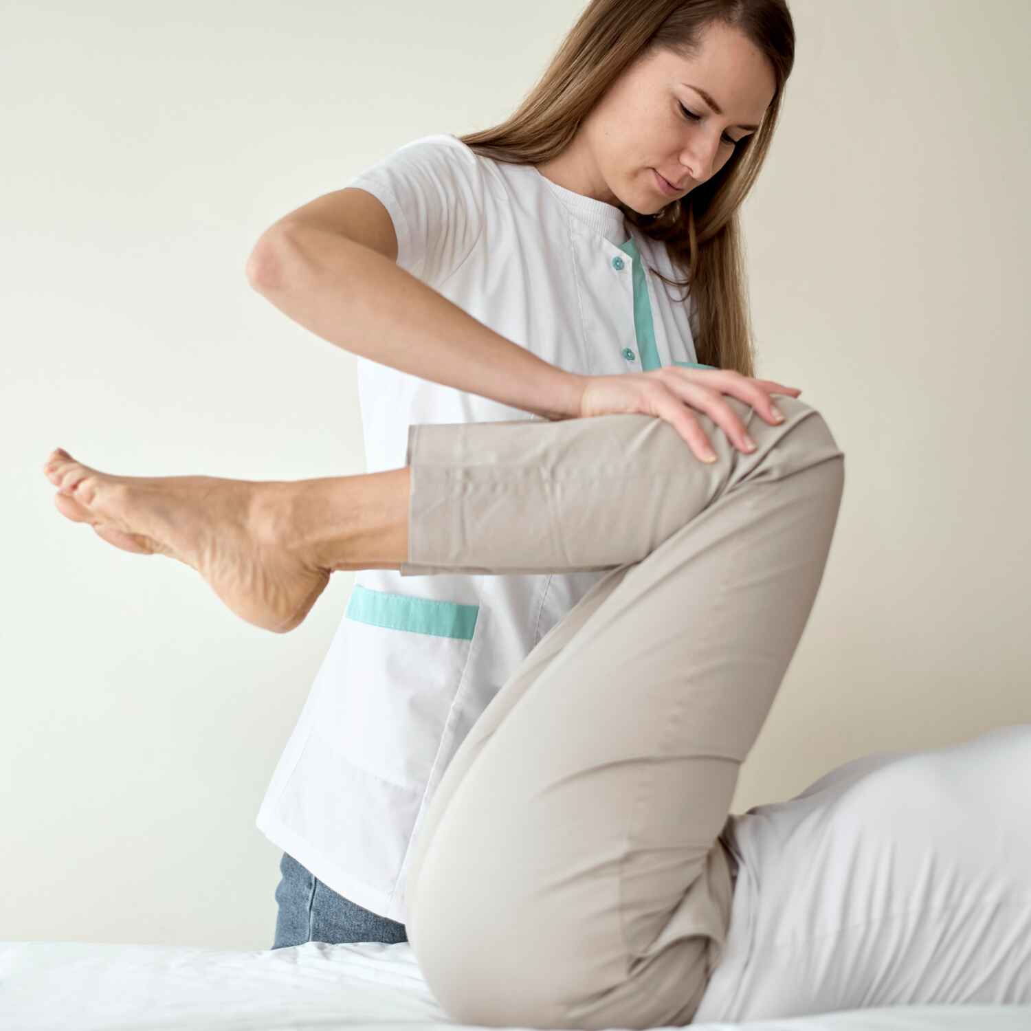
Overview
The meniscus, which serves as a shock absorber between the femur and tibia, is commonly injured in traumatic knee events. Meniscal tears can occur through axial loading, twisting, or bending movements.
Meniscal Injuries
Traumatic ligament injuries commonly affect the anterior cruciate ligament (ACL), posterior cruciate ligament (PCL), medial collateral ligament (MCL), and lateral collateral ligament (LCL). These injuries often result from high-impact trauma, such as during sports activities.
Meniscectomy
In cases of severe meniscal tears, where the meniscus cannot be repaired, partial meniscectomy is performed to remove the damaged portion. This helps reduce pain and prevents further damage to the knee joint.
Meniscal Repair
If the tear is in a region where healing is possible (such as the outer 1/3 of the meniscus, which has a blood supply), a meniscal repair can be performed arthroscopically. This procedure aims to preserve the meniscus, preventing the long-term risk of osteoarthritis.
Meniscal Transplantation
In cases where the meniscus is severely damaged or removed, a meniscal allograft transplant may be performed to restore function and reduce the risk of arthritis.

Advantages of Arthroscopic Surgery
Minimally Invasive
Smaller incisions reduce the risk of infection, blood loss, and postoperative pain.
Faster Recovery
The minimally invasive nature of the procedure allows for a quicker return to activity and less postoperative downtime.
Autologous Chondrocyte Implantation (ACI)
Autologous Chondrocyte Implantation (ACI) involves harvesting healthy cartilage cells from the patient, culturing them, and then implanting them into the defect site.
Better Visualization
High-definition cameras and specialized instruments offer superior visualization of internal knee structures, allowing for precise and accurate treatment.
Less Scarring
Smaller incisions reduce scarring compared to traditional open surgery.
Surgical Management Of Recurrent Dislocation Of Patella.
Recurrent patellar dislocation occurs when the patella (kneecap) repeatedly dislocates or subluxate (partially dislocates) from its normal position in the femoral groove. This condition is often the result of a combination of anatomical, functional, and environmental factors, such as a shallow trochlear groove, patellar malt racking, ligamentous laxity, or previous traumatic injury. The management of recurrent patellar dislocation is typically aimed at stabilizing the patella and preventing further episodes.
Surgical Approaches
Medial Patellofemoral Ligament (MPFL) Reconstruction
Indication
MPFL reconstruction is one of the most common surgical procedures for recurrent patellar dislocation, especially when the medial patellofemoral ligament (MPFL), a critical stabilizer of the patella, is torn or lax.
Procedure
The MPFL is reconstructed using a tendon graft (usually the gracilis or hamstring tendon), which is positioned to restore the normal function of the ligament. The graft is anchored to the femur in a position that mimics the original ligament's attachment.
Outcome
This procedure is highly effective in reducing the risk of recurrent dislocation in most patients, with good long-term functional outcomes.
Tracheoplasty
Indication
Trochleoplasty is considered when the underlying issue is a shallow or dysplastic trochlear groove that fails to adequately support the patella. This can be seen in patients with a high Q-angle or abnormal patellar tracking.
Procedure
The surgeon reshapes the femoral trochlear groove to create a deeper and more stable track for the patella to sit in. This can involve cartilage and bone work.
Outcome
While it can be very effective in patients with trochlear dysplasia, it is a more complex procedure and carries a higher risk of complications compared to other techniques.
Tibial Tubercle Transfer (Tuberosity Osteotomy)
Indication
Tibial tubercle transfer is used in patients with a high-riding patella or malalignment of the patellar tendon relative to the femoral groove. It helps to realign the patella and restore normal tracking.
Procedure
The tibial tubercle is moved to a more favorable location on the tibia, usually medially and proximally, in order to better align the patella with the femoral groove.
Outcome
This surgery is effective for addressing patellar maltracking caused by anatomical malalignment, particularly in cases where MPFL reconstruction alone is insufficient.
Lateral Release
Indication
A lateral release is sometimes performed when tight lateral structures (such as the lateral retinaculum) contribute to abnormal patellar tracking or when the patella is laterally subluxing.
Procedure
The surgeon releases or cuts the tight lateral structures around the patella, allowing it to move more freely and reducing the lateral force.
Outcome
This procedure is often used in combination with other surgeries and is typically performed in cases where there is excessive lateral pull on the patella. It may not be as effective alone in preventing dislocations.
Realignment of the Patella
Indication
This can include both tibial tubercle transfer and other soft tissue procedures designed to correct patellar alignment.
Procedure
The aim is to restore proper alignment of the patella in the femoral groove, ensuring that it tracks properly during knee motion.
Outcome
This is a useful approach when there is significant maltracking of the patella, and other less invasive options have failed.